Fertility Tests – Female
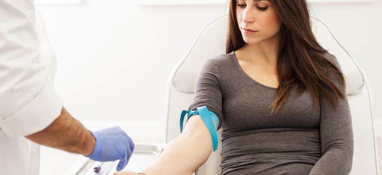
Blood Investigations are done to assess egg reserve and rule out any major hormonal ,medical or infective condition which may be contraindicatory to treatment or pregnancy. Thalasemia is done so that it is not passed on to the baby and Rubella IgG is tested so that the lady is adequately immune.
- FSH, LH, Serum estradiol, AMH
- Thyroid profile
- Prolactin
- Blood Sugar
- Thalasemia
- Rubella IgG
- Viral markers
Radiological Assessment
Hysterosalpingography (HSG) and sonosalpingography are both diagnostic imaging techniques used to evaluate the female reproductive system, specifically the uterus and fallopian tubes. However, there are differences in how they are performed and the imaging modalities used:
- Hysterosalpingography (HSG) : HSG is a radiographic procedure that uses X-rays to visualize the uterine cavity and fallopian tubes. During the procedure, a contrast dye is injected into the uterus through the cervix, and X-ray images are taken as the dye fills the uterus and flows through the fallopian tubes. This helps identify any abnormalities, such as uterine malformations, blockages, or tubal patency issues like Hydrosalpinges. HSG can be useful in diagnosing causes of infertility or recurrent miscarriages.
- Sonosalpingography (SSG) : Sonosalpingography is a similar procedure to HSG, but instead of using X-rays, it utilizes ultrasound imaging to visualize the reproductive organs. In sonosalpingography, a saline solution or a contrast agent is injected into the uterus through the cervix. The fluid fills the uterus and flows through the fallopian tubes while ultrasound imaging is used to monitor the process in real time. This allows the examiner to assess the shape and condition of the uterus and detect any abnormalities or blockages.
The choice between HSG and sonosalpingography may depend on several factors, including the availability of imaging equipment, patient preference, and the expertise of the healthcare provider. Both procedures have their advantages and limitations, and the specific circumstances of each case will determine which one is more appropriate.
It’s important to consult with a healthcare professional to determine the most suitable diagnostic imaging technique based on your specific needs and medical history. We perform SSG at our centre under deep sedation as without it , it can be a painful procedure.
The role of Transvaginal Ultrasound (TVS) in fertility is very important.
For a pelvic ultrasound one must have an empty bladder and one must be prepared for vaginal probe insertion. For women who have non-consummated marriage or do not want the probe to be inserted, we have the transabdominal scan done with full bladder. However transabdominal scan never gives as good a picture as transvaginal scans.
The Transvaginal Ultrasound gives an image of the ovaries, uterus and pelvic structures. It states the total no. of antral follicle count which indirectly reflects the ovarian reserve and helps us differentiate between low, normal and poly cystic ovaries. It also gives an idea of the uterus whether there are fibroids, polyps, and about the endometrial thickness. Along with this we come to know about the pelvic structures, cysts, hydrosalpinx and adhesions.
Pelvis ultrasound is usually done initially when the cycle starts, in which we see thin endometrium and the ovaries are quiet with no follicle/cyst formation. If the endometrium is thick or we see cyst formation, then blood estrogen levels are done to check whether that cycle is good enough for stimulation or not. The next scan is done depending on the procedure. TVS can be done in both 2D and 3D. 3D gives more clarity and is useful in uterine defects, hydrosalpinges etc. When we are trying to evaluate a lady for female fertility, we do another scan on the 15-18 day of the menstrual cycle and this gives a very important info about the endometrial thickness, which is key for the success of fertility treatment. A good endometrial thickness is between 8-10mm on day 15-18.
MRI – This is an advanced test which is used to find the exact location and size of fibroid, adenomyosis , endometriotic cysts and hydrosalpinges.
Diagnostic Laparoscopy and Hysteroscopy
Hysteroscopy is a procedure where we put a camera into the uterus and check whether there are any fibroids, polyps or adhesions.
Laparoscopy is a procedure where we put a telescope into the abdomen and check the outer part of the uterus, tubes and ovaries with our own eyes.

